ANATOMY AND PHYSIOLOGY OF EXTERNAL EAR
INTRODUCTION
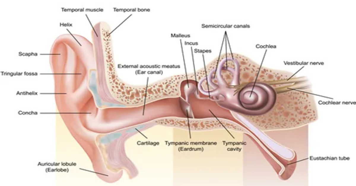
Auditory system comprises the ears and its connections to and within the central nervous system. Anatomically the auditory system can be divided into the outer, middle, and ears; the auditory nerve; and the central auditory pathways
The outer ear is made up of the pinna (auricle) and ear canal(external auditory meatus). The ear drum (tympanic membrane) separates outer and middle ear and is generally considered to be part of middle ear. The middle ear also includes the tympanic (middle ear) cavity; the osscicular chain with its associated muscles ,tendons, and ligaments; and the Eustachian (auditory)tube. The inner ear begins at the oval window. It includes the sensory organsof hearing (the cochlea) and of balance (semicircular canals,utricle, and saccule)
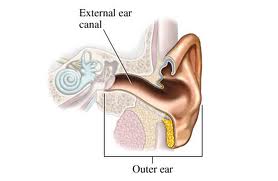
EXTERNAL EAR
The external ear is composed of the pinna and the ear canal, ending at the tympanic membrane.
PINNA
Pinna is a latin term for wing or projecting part of ear lying outside head. It is also called as Auricle. In latin term ‘aural’ pertains to ear. It’s the externally visible aspect of ear, irregularly shaped ovoid of highly variable size.It folds at the side of head at 30degree,situated posteriorly, superiorly and interiorly.It is composed of skin covered elastic cartilage.It contains some undifferentiated muscles and number of extrinsic muscles which are vestigeal in humans.
SURFACE ANATOMY OF PINNA
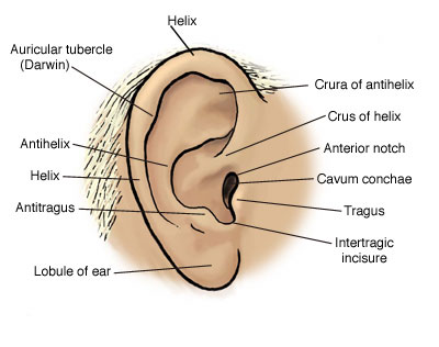
The major landmarks of the pinna are highlighted in the above figure. In the following the description about these landmarks are given.
Concha-It is the entrance to the ear canal and at the bottom , it’s large cup shaped depression.
Tragus– Concha is partially covered by a posteriorly directed projecting or ridge called tragus.
Anti-tragus-To the opposite of tragus,a smaller ridge forming an inferior boundary of concha,is called the anti-tragus.
Inter-tragal incisures– The tragus and anti tragus are separated by a notch called inter tragal incisure.
Helix –The ridged rim along most of the perimeter of the pinna.
Crus of helix-Starting at the very top of pinna, the helix courses anteriorly and downward, making a hook like turn in the posterior direction to form a shelf above concha
Lobule-Helix courses downward along the posterior perimeter reaching the ear lobe or lobule.
Darwin’s tubercule– A frequently found variation of helix near the tip posteriorly, is a thickened portion called Darwin’s tubercule.
Scaphoid fossa -a depression between helix and anti helix is called ‘scaphoid fossa’ or ‘boat shaped ditch’
Cymba-Superior part of concha divided by crus,is called cymba.
Cave-Inferior part of concha divided by crus is called cave.
INTERNAL STRUCTURE OF PINNA
The core of the auricle consists of a fibrous cartilage,shaped roughly similar to ear. Medially the auricular cartilage is continuous with the cartilagenous skeleton of the ear canal. The anterior, superior and posterior auricular muscles which are attached to the auricle are vestigeal. Some small intrinsic muscles extend from one part of auricle to other are also non-functional.
VARIABILITY
The auricles are highly variable in their shape, to the extent they have been used for purposes of identification much as fingerprints have been used.
EXTERNAL AUDITORY MEATUS
Communication between the middle ear & the inner ear and the external environment is provided by “external auditory meatus” or simply “ear canal”. It leads from the concha to eardrum or tympanic membrane. It varies both in size and shape. The outer 1/3rd portion is cartilagenous, remaining 2/3rd is bony. The junction between bony and cartilagenous portion is called ‘isthmus’.
CONFIGURATION
EAM is a curved and irregularly shaped tube called ‘S’ shaped. The course of ear canal is more horizontal in infants and small children than adults. The diameter is largest at the ‘Auricular (external) orifice, gradually smaller toward isthmus, expands again only to decrease in size just before the tympanic membrane.
According to Wever & Lawrence,1982, at the entrance, the diameter is 0.7cm,average horizontal diameter is 0.65cm, mean vertical diameter is 0.9cm. According to Donaldson & Miller,1973, the length of the canal is not uniform. Posterosuperiorly, the length is approximately 2.5 cm and inferoanteriorly, its 3.1cm. According to Gelfand, in average, the height is 9 mm, width is 6.5 mm, length is 2.5 to 3.5 cm.
LINING OF THE MEATUS
The skin lining is closely adherent to periosteum and perichondrium of supportive skeleton. It forms the outer layer of ear drum’s membrane. The canal’s skin is thicker in the cartilagenous segment than in bony part. The bony portion of canal is derived from
- The tympanic part of temporal bone which forms the floor and anterior wall as well as inferoposterior wall.
- The squamous part,make up the roof and part of posterior wall.
- The condyle of mandible which contributes to the inferoanterior wall t temporomandibular joint.
The cartilagenous portion contains hairs, plentiful distribution of sebaceous (oil) & ceruminous (wax) glands.
PHYSIOLOGY OF EXTERNAL EAR
FUCTION OF PINNA
- Non-auditory function:
- Protective – protects ear canal from foreign body (tragus covers ear canal opening)
- Cosmetic purpose
- Auditory function:
i)Collecting/Funneling sounds to the ear canal
ii) Enhancing Localization
iii) Resonance property of Concha
i) Funneling sounds to the ear canal
Acoustically, the pinna without the concha is termed the pinna flange. It has minimal acoustic effect. The pinna with its ridges, grooves and dished out regions, is an excellent funnel for information directed toward the head from front or side, although less effective for sound arising from behind the head.
The collection of sounds or funneling function is not significant for human pinna. This was examined in two different ways (Békésy and Rosenblith, 1958)
- The depressions in the pinna was filled with wax and by doing this there was no reduction in hearing sensitivity.
- Insertion of tube to avoid the function of pinna, which showed no difference in the hearing sensitivity.
Thus pinna has no role in collection of sounds to the EAC.
ii) Enhancing Localization
The pinna’s greatest contribution to hearing is probably in sound source localization. The obvious example occurs when a pet turns its ears towards a ringing doorbell. This action is impossible for man, whose extrinsic ear muscles are vestigial. However, head turning in both humans and animals orients the ears relative to the stimulus, which, along with difference in the intensity and time of arrival of a sound at the two ears permits accurate localisation.
The pinna contributes to localization in man, particularly when listening with only one ear (monaural localisation) and when the sound source is in the median plane of the head rather than off to one side. Pinna effects are important in these two situations because monaural hearing precludes the use of the inter-aural difference available during binaural hearing, and because these inter-aural differences are minimized in the median plane, where the sound source is equidistant from both ears.
The depressions and ridges of the pinna contribute to median plane localization, (as evidenced by increases in the number of localization errors made when the various depressions were filled). The contribution of the pinna to localization is due to variations in the stimulus spectrum caused by the structure of the pinna.
There are two hypotheses to explain how pinna helps in localization:
- Filter action
Blauert (1967) suggested that the pinna acts as a filter that attenuates or passes frequencies depending on the direction (front sounds are enhanced and sounds from back are attenuated)
- Time delay
Batteau (1967) showed that sounds reflected from the structure of the pinna cause time delays between the reflected and direct source on the order of under 300 micro sec. Since these very small delays are detectable, they may be used in median plane and monoral localization (the difference between actual signal and reflected signal is different for different directions which helps in localisation).
Frequencies over 4000 Hz are particularly important in these effects.
Types of localization are:
- Horizontal localization
Horizontal localization is made possible by differences in Interaural time and intensity differences.
- Interaural time difference
Sound will arrive at the ear closer to the source (near ear) before they arrive at the other ear (far ear).The resulting difference in time at the two ears is called the Interaural time difference.
Klumpp and Eady (1956) determined the minimal time delay permitting listeners to lateralize a tone correctly to the side of the leading ear 75% of time. A minimum of about 10usec found for 1 KHz tone.
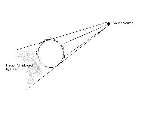
Figure: The distance from the sound source to each ear is different therefore the sound is delayed longer in reaching the far ear. This helps humans perceive the spatial location of the sound.
- Interaural Intensity difference / Interaural Level difference:
Reduction in the amount of intensity when the signal is reaching the far ear side.
The ear that is farther from the sound source will often not have a direct path for the sound wave to follow because the head is in the way. This is shown in the figure above. Thus the sound wave reaching the far ear will be attenuated compared to the near ear, creating an inter-aural level difference between the ears (ILD).
This is due to the head shadow effect – A head shadow occurs when the head is between the sound source and the ear being investigated (significant for frequencies over 1500 to 2000 Hz).
ILDs are not linear with frequency. Low frequencies do not appear to be shadowed much by the head because they have a wavelength which is greater than the diameter of the head. Frequencies above 1500 Hz have a wavelength smaller than the diameter of the head, and can be attenuated a great deal. It follows then that high frequency components are much more important to us when evaluating ILDs because they are most affected by the head shadowing resulting from the location of the sound source.
Lord Rayleigh (1907) proposed “duplex theory” which considered that high frequencies are located laterally by the inter-aural intensity differences whereas low frequencies are located laterally by the inter-aural phase differences (time).
- Vertical localization:
In the medial plane it is made possible by reflections and resonances that occur within the pinna prior to sound entering the ear canal. Reflection causes cancellation and hence spectral peaks and notches. Resulting cues to localization in the mid sagital vertical plane are all above 5 kHz because only in this high frequency region that the wavelength of the sound is small enough, compared to the size of the pinna for necessary reflection and resonance to occur. People can detect changes in vertical angle as small as 3o.
- Front back localization
The pinna boost high frequency sounds when they arrive from front, but attenuates them when they arrive from rear. Sound during the first few milliseconds of a signal has a particularly strong influence on perceived duration.
- Monaural localization:
Localization using monaural cues in which stimulus is presented to one ear alone.
Mostly depends on the filtering effects of the external structures. These structures include head, torso, shoulders, and outer ear or pinna. Sounds are frequency filtered specifically depending on the angle from which they strike the various external filters. The most significant filtering cue for this type of localization is the pinna notch, the notch filtering effect resulting from the destructive interference of wave reflected from the outer ear. In order to enhance filtering mechanism, many animals have evolved large specially shaped ears. Many also have the ability to turn the outer ear at will which allow for better sound localization. Bats and barn awls are paragons for monaural localization in the animal kingdom.
Morimoto (2001) investigated the contribution of each pinna to the perception of vertical angle. Tests measured localization of the vertical angle in five planes parallel to the median plane. In the localization tests, the pinna cavities of one or both ears were occluded. Results showed that pinna cavities of both the near and far ears play a role in determining the perceived vertical angle of a sound source in any plane, including the median plane. As a sound source shifts laterally away from the median plane, the contribution of the near ear increases and, conversely, that of the far ear decreases. For saggital planes at azimuths greater than 60 degrees from midline, the far ear no longer contributes measurably to the determination of vertical angle.
The gain produced by the pinna is not very large, approximately 3 dB at 4 KHz. Because of its shape, the effect of the pinna varies considerably with the angle of incidence of the sound.
iii) Resonance property of Concha:
Concha produces a gain of approximately 10dB at 4-5 kHz but a loss of approximately 5dB at 10 kHz. The concha functions as a cavity resonator. The frequency of resonance depends on the diameter and the depth of the concha. Average concha resonates around 4700 Hz. The deeper and larger the concha lower is the resonant frequency.
FUNCTIONS OF EAR CANAL
Non-auditory functions:
This includes the protection of the tympanic membrane and deeper structures from injury and the maintenance of a clear and disease free passage through which sound can be conducted to the tympanic membrane.
- Protection Mechanism:
- Anatomical structure:
The protective function of the external ear canal is directly related to its physical shape and properties.
The shape (S shaped course of the EAC) and its depth of the EAC and rigidity of its walls provide protection to the tympanic membrane from direct injury.
The deeper, narrower and more tortuous the canal, less will be the chances of direct injury.
ii) The epidermal covering:
- The epidermal layer provides the barrier that protects the underlying tissues from the potentially harmful effects of the surrounding environment (i.e. drying, wetting and chemical damage).
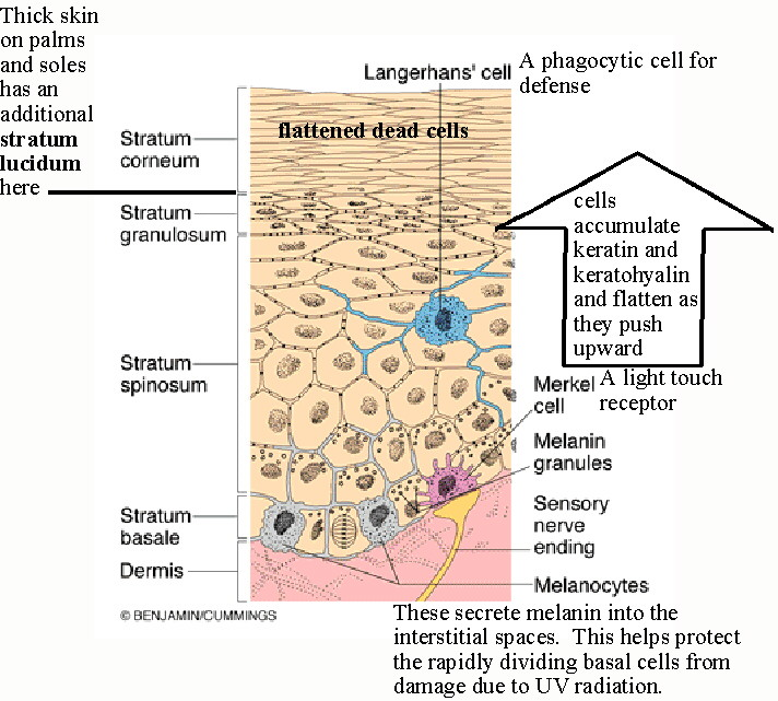
- This protective mechanism is provided by the most superficial layer, the stratum corneum which consists of layers of dead keratinocytes
- As the cells of the stratum corneum have terminally differentiated, they are incapable of either self-regeneration or repair.
- The integrity of the stratum corneum must therefore be maintained by the continous production of keratinocytes from the deeper layers of the epidermis that have not yet terminally differentiated, i.e., the cells of the stratum basale and stratum spinosum.
- These replacement cells that arise in the stratum basale undergo differentiation as they rise through stratum granulosum.
- At the upper layer of stratum granulosum, these rising cells undergo terminal differentiation into keratinocytes joining the deepest layer of the stratum corneum.
- When the keratinocytes reach the upper surface of the stratum corneum they are continuously shed from the surface of the body. This process is known as desquamation.
Acidic ph of the skin surface is another microbial deterrent (6.5-6.8) which is significantly below the optimal ph (7 or higher) for the growth of pathogenic bacteria.
- Hairs
- Hairs may be involved in defense.
- Hairs are found in the outer cartilaginous part and in the posterior-superior part of the bony portion of the EAC.
- Hairs point outward so that these outward facing hairs in the cartilaginous part of the ear canal hinder the entry of the invaders whether it is animate (such as an insect) or inanimate.
- The hairs are short and the most medial hairs lie at a very oblique angle, almost flat, against the canal wall whereas laterally they become more upright and therefore more obstructive to anything entering the canal.
- Trap-like arrangement of the hairs in the canal, and the nature of the surface of individual hairs may contribute to the one-way system in the canal. This arrangement may facilitate movement from the root toward the tip of the hair but not in the opposite direction.
- Wax
- Wax or cerumen is a combination of desquamated keratinocytes and the secretions from the glands of the superficial part of the external ear canal.
- There are two types of secretory Glands in the EAM
- Ceruminous
- Sebaceous
- Ceruminous glands are a type of apocrine gland.
- Sebaceous glands are closely associated with hair follicles and are simple or branched alveolar glands, which produces holocrine fatty secretion of triglycerides and wax esters that empties into the canal at the base of the hair follicles.
- Sebum has a slight antibacterial and antifungal properties, as well as waterproofing effect on the surface of the epithelium.
Cerumen glands and cerumen secretion
- The ceruminous glands number between 1000 and 2000 in normal ear canal and have a distribution similar to the hairs of the ear canal
- Ceruminous glands are tubular glands with ducts that may open either directly into the base of the hair follicle superficial to sebaceous gland duct.
- Contraction of the myoepithelial cells causes the contents of the ceruminous glands to be secreted. (Perry et al)
- Ceruminous glands are stimulated to adrenergic stimulations such as locally or systemically injected adrenaline or pain and anxiety.
- Local pressure on the skin and movements such as clenching of teeth also stimulated secretion.
- These secretions coat the skin and superficial part of the canal.
Functions of cerumen / Wax
- Acts as water repellent
- Entraps dust, hair follicles , and insects
- The primary protective function of cerumen is repelling insects. (Perry 1957)
- The physical stickiness of these secretions may have some protective function in that objects coming into contact with it are more likely to stick to the skin and hairs of the superficial canal.
Physiology of cerumen impaction
- Cerumen impaction is the result of the failure in the breakup or separation of the individual keratinocytes which normally occurs in the superficial external auditory canal.
- At present little is known about mechanism, whereby the continuously desquamating layer of deep canal stratum corneum is broken up and spontaneously expelled from the ear canal. (Robinson et al 1990)
- It is found that the chemical compound that facilitates this detachment is not known and is called as keratinocytes attachment destroying substance (KADS). (Robinson et .al 1990)
Causes for impaction:
- Thicker and longer hair can lead to impaction.
- Also because of obstruction due to hearing aids.
- Using cotton tipped swabs actually pushes the wax inside. Rather than extracting the wax, it impacts it deeper into the EAC. (Johnson & Hawke 1988)
Self-cleansing function:
The epidermis lining the EAC is in a unique situation; because of its location, the epidermis is not subject to the usual surface contact (friction) that normally removes those keratinocytes that have desquamated from the surface of the stratum corneum elsewhere in the body.
This means that an alternative mechanism (self cleansing) exists for the removal of dead cells in the EAC.
Epithelial migration
- The purpose of the migratory phenomenon is clearly to keep the canal free of debris. This phenomenon was first documented by Blake(1882)
- When skin is migrating of the tympanic membrane and along the EAC, cells have to be generated to replace those leaning. In the normal ear canal the surface of the skin moves laterally from the medial end of the canal
- The rate of migration in the external ear canal in humans is similar to the nail growth.
- Litton stated that the generation center in the humans would be expected in the region of the umbo. Migration of skin is probably a combination of active migration (deep stratified layer of the skin) and passive migration (Keratin dispersion).
- There are two types of migration with respect to umbo and manubrium.
- The lateral movement of epithelium with respect to umbo is called radialpattern of movement.(found in 80%)
- The lateral movement of epithelium with respect to manubrium is called horizontal pattern of movement.
- The emigrating epidermis has to come to a halt at some point in the EAC. This differentiates into the dead cells of the stratum corneum followed by the detachment (desquamation) from the skin surface.
Auditory Functions:
- Resonance properties of EAC
- Role of Ear Canal and Pinna combined
- Azimuth
- Resonance effect of the ear canal
- External auditory meatus is closed at one end by tympanic membrane and open at the other end.
- In such a tube resonation occurs when the length of the tube is 1\4th the wavelength of the applied sound (quarter wavelength theory)
L= l/4 →→→ l= 4L.
- Weiner and Ross (1946) found the average length of meatus to be 2.3cm.
l=4*2.3= 9.2cm= 0.092m.
f= v/l= 330/0.092= 3.6kHz.
Thus the resonant frequency of the EAC is around 3.6 kHz (2.5 to3.6 kHz)
- In children the resonant frequency will be higher (as the length of the EAC is short)
- Thus shorter the length of the EAC greater will be the resonant frequency.
- The gain given by the ear canal at resonant frequency is around 17 dB if the walls of the ear canal are rigid. But as the tube is not straight and also covered with skin, it absorbs some sound energy and it increases the sound signal approximately by 15 dB.
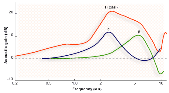
- Role of Ear Canal and Pinna combined:
When pinna flange and concha combined with the ear canal, sound pressure gain curve is slightly different.
The concha resonance blends with the ear canal resonance producing a composite curve with two peaks one at 2700 Hz and the other at near 5000 Hz.
Weiner and Ross (1946) – measured sound pressure distribution in the ear canal from a free field sound presented at three angles of incidence: 00, 450 and 900 in the horizontal plane. The pressure distribution in the ear canal at various frequencies varied, and the most obvious pressure gain was reported in the region of 2 – 4 KHz (10 – 15 dB) with a maximum increment of 17 to 22 dB at 3 KHz. Hearing Aid Center in Kolkata.The important findings of the study are:
- The outer ear that is concha and ear canal acts as a resonator and the amount of resonance provided is up to 20 dB between frequencies 2 to 5 KHz.
- The sound source has a differential effect on the amount of pressure developed in the canal across frequencies.
- The gain for a 450 angle being the largest.
Azimuth:
Azimuth is the angle at which the ear is oriented towards the sound source.
The SPL value will differ depending on the azimuth.
This is mainly because of two factors:
- Reflection and diffraction of the sounds from head and other body parts.
- Head shadow effect
Effect of the external ear on Bone conduction thresholds
Tondorf (1968) provided the explanation for the external canal component of bone conduction in order to account for the occlusion effect, in which low frequency bone-conducted signals are perceived as louder when the ear canal is occluded than when it is unoccluded. When a signal is introduced to a skull through an oscillator, the osseous and cartilaginous walls of the ear canal vibrate along with the skull.
- As a result, sound energy is radiated into the space within the canal.
- Some amount of energy is transmitted through the ear canal to the cochlea.
- If the ear canal is unoccluded, the ear canal acts as a high-pass filter.
- If the canal is occluded, the high-pass filter effect is eliminated, so low-frequency transmission is improved by about 10dB in 250-2000Hz region. Hearing Aid Center in Kolkata
References:
Gelfand, S.A. (1988). Hearing- An introduction to psychological and physiological acoustics. 3 rd Ed. New York: Mared Dkkr.
Moller, A.R. (1983). Auditory physiology. New York: Academic press.
Pickles, J.O. (1992). An introduction to physiology of hearing. New York: Academic press.
Yost, S.A. (1994). Fundamentals of hearing: An introduction. 3 rd edition. San Diego: Academic Press, Inc.
Zemlin, W.R. (1998). -Speech and hearing science: Anatomy and Physiology. 4 th edition. Boston: Allyn and Bacon.
Presented by
Soudamini Rout(MASLP-1